Carotid Dissection
Carotid artery dissection is a condition whereby the layers of the carotid artery are spontaneously separated - this potentially compromises blood flow to certain areas of the brain and can lead to a stroke. It can occur extracranially or intracranially and can lead to subarachnoid hemorrhage or brain ischemia. It is the most common cause of strokes in younger patients and the importance of making a timely diagnosis is paramount to minimise potential morbidity and mortality of the disease.
There is huge variation in the presenting signs and symptoms of carotid artery dissection, making it extremely difficult to diagnose on title presentation.
Etiology
The tear can be spontaneous or caused by trauma. The intramural hematoma causes stenosis and eventual thrombus formation. Blunt trauma can be significant such as a RTC or can seen minimal (chiropractic manipulation) - it can also occur with penetrating trauma. A RTC where there is rapid deceleration with simultaneous neck hyperextension and rotation may lead to an intimal tear of the carotid artery. Idiopathic dissection is the most common cause of spontaneous dissections in which a family history is present:
Ehlers-Danlos
Fibromuscular dysplasia
Other connective tissue disorders
An elongated styloid process, known as Eagle syndrome, can also cause a spontaneous internal carotid artery dissection
Epidemiology
Carotid artery dissections occur in all age groups. They account for 2.5% of all strokes - it is a common cause of stroke in patients under the age of 40. In all young patients, 20% of strokes are caused by carotid artery dissection. The median age is the mid-40s, with a slightly higher incidence in males versus females. The incidence has been found to be higher during autumn, with literature suggesting the peak incidence occurs in October. It is proposed that weather-related factors affect blood pressure, coagulation, blood flow, physical activities, and diet. Air pollution also may contribute to vascular changes, leading to carotid artery dissection. Migraines have been described to be an independent risk factor for dissection.
Pathophysiology
A sudden tear in the intimal layer of the carotid artery occurs due to the factors listed above - it may be due to trauma, or they can be spontaneous. This tear allows blood to flow into the intimal layer of the vessel and allows a hematoma to form within the blood vessel wall - a false lumen.
As blood enters the false lumen, it causes stenosis which may lead to complete occlusion of the carotid artery. This is a dynamic process that may lead to dilation of the artery depending on where the hematoma develops; either towards the intima or adventitia. This may lead to a stroke from a complete vascular occlusion at the site of dissection.
History and presentation
Presentation can range from an asymptomatic patient to one who present with an acute stroke. Classical, the patient will present with headache, facial, or eye pain and neck pain. The pain when present is usually on the ipsilateral side to the dissection. Horner syndrome may be present if a hematoma of the cervical artery compresses the adjacent sympathetic nerve fibres. When stroke-like symptoms are present with these symptoms, it can make the diagnosis somewhat less difficult.
Neurological defects should prompt one to consider this diagnosis especially if the patient is young and there is recent trauma. Recent trauma to the neck with anterior neck pain is a potential clue alongside tenderness. A bruit over the carotid may be heart and a expanding hematoma may be present if trauma has occurred.
Investigations and diagnosis
If cervical dissection is suspected, many diagnostic modalities may confirm the diagnosis. The initial and least invasive screening tool is a carotid ultrasound; its sensitivity is not as good as CT angiogram and it does not allow for imaging of the intracranial vessels. A CT angiogram is more commonly ordered these days as it can be done concurrently with a brain CT looking for acute stroke or intracranial bleed.
A flame sign is a classical sign that can be seen; it is due to tapering of the carotid artery. MRI and MRA are reasonable alternatives to CT if the patient has contraindications to CT angiogram.
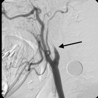
According to ESO guidelines, a carotid artery dissection should be split into extracranial artery dissection (EDA) and intracranial artery dissection (IAD). EAD refers to the dissection of a cervical carotid or vertebral artery radiologically confirmed by the presence of a mural haematoma, a dissecting aneurysm, a long tapering stenosis, an intimal flap, a double lumen or an occlusion >2 cm above the carotid bifurcation revealing a dissecting aneurysm and/or a long tapering stenosis after recanalization.
Treatment and management
Treatment of a cervical dissection depends on multiple factors - cause, acute stroke or IAD/EAD. If there are no contraindications, antiplatelets may be used, or more commonly, systemic anticoagulation may be used to minimise the risk of a stroke. There is a theoretical concern that thrombolysis may increase the risk of enlargement of an intramural hematoma in the dissected artery and thus impair cerebral hemodynamics, or promote dissecting aneurysm formation or vessel rupture. An EOS systemic review identified no randomised data on the efficacy and safety of IVT in patients with EAD/IAD.
Endovascular stenting may be performed on some of these patients, especially if there are contraindications to anticoagulation or if medical management fails.
Differential Diagnosis
Acute hypoglycaemia
Carbon monoxide toxicity
Cervical fracture
Cluster headache
Hemorrhagic stroke
Herpes simplex
Herpes zoster
Ischemic stroke
Migrane
Neck trauma
Retinal artery occlusion
Retinal vein occlusion
SAH
Tension headache
TIA
Vertebral artery dissection