Superior vena cava obstruction
Superior vena cava syndrome (SVCS) is a collection of clinical signs and symptoms that result from either partial or complete obstruction of blood flow through the SVC - it most commonly occurs as a result of thrombus formation or tumour infiltration of the vessel wall. The SVC is formed by the junction of the left and right brachiocephalic veins and returns blood from the head, neck, upper limbs and torso to the heart.
Aetiology
SVCS saw a large decrease throughout the 20th century - most cases now are due to mediastinal malignancies, primarily small cell bronchogenic carcinoma. It can also occur due to non-Hodjkin's lymphoma, followed by metastatic tumours. Benign/non-malignant cases of SVCS compromises at least 40% of cases.
Iatrogenic thrombus formation or SVC stenosis is a growing aetiology due to pacemaker wires and semipermanent intravascular catheters used for hemodialysis, long-term antibiotics, or chemotherapy.
Epidemiology
25 years ago, malignant causes accounted for >90% of all cases, but there has been an increase in benign causes due to the use of intravascular devices as mentioned above. SVC syndrome occurs in approximately 15,000 people in the US every year. Of these, malignancy compromises 65% of cases, most commonly lung and non-Hodgkins lymphoma. Malignant causes are more common in middle-aged to older adult males, while benign causes are equally distributed across both sex and gender. Infectious causes such as syphilitic aortic aneurysm and tuberculosis accounted for the majority of non-malignant cases in the past, but is now a rare occurrence especially in developed countries.
Pathophysiology
The SVC is part of a low-pressure venous system which contains thin walls that are susceptible to damage; large thick walls are not required due to low pressures. The mechanisms that can cause damage to the SVC can be divided into:
Compromised vessel anatomy
Impaired venous flow
Diminished vessel wall integrity
In patients with SVCS, these mechanism often co-exist. In a patient presenting with a mediastinal mass, compression and obstruction of the SVC by the mass is the most common. However, there are non-malignant causes such as dilation of the overlying aorta.
Venous thrombus SVCS is increasing as mentioned above. This can also cause venous wall inflammation, fibrosis and eventual thrombus that leads to stenosis of the vessel itself.
History and presentation
The symptoms that SVCS causes is due to the collateral vascular network that serves to divert blood to the lower body, where it is returned to the heart via the inferior vena cava (IVC), azygos vein and the intercostals. The findings are related to venous congestion and the resultant elevation in venous pressure seen in the upper body. A cardiac exam is sufficient to rule out cardiogenic origins of the patient symptoms - such as increased pressure in the SVC secondary to right sided heart failure or tricuspid valvulopathies.
The most common presenting symptoms are:
Face/neck swelling
Distended neck veins
Cough
Dyspnoea
Orthopnoea
Upper extremity swelling
Distended collateral chest vein
Conjunctival suffusion
Enlargement of the conjunctival vessels
Other symptoms include stridor, hoarseness, dysphagia, pleural effusion. It may also present with head plethora - headache; nausea; lightheadedness; syncope; change in vision; altered mental status; upper body oedema; cyanosis; papilledema; stupor and coma.
Rarer but serious clinical consequences include:
Cerebral oedema
Upper respiratory compromise secondary to oedema of the larynx and pharynx
Investigations and diagnosis
According to BMJ best practice, 1st line imaging investigations to order are:
CXR : Widened mediastinum or mass lesion in the lung
CT Chest : Full or partial obstruction; development of collateral vessels; shows location, severity and associated pathology
MRI Chest
Ultrasound of upper body and vasculature (screening for thrombus)
Patients with high clinical suspicion for SVC should undergo imaging of the upper body and vasculature. Ultrasound of the jugular, subclavian and innominate veins can help identify a thrombus within the vessel lumen.
CT of the chest for the presence of collateral vessels is associated with a diagnostic sensitivity of 96% and a specificity of 92%.
CT venography is accepted as the gold standard for visualizing and diagnosing a venous obstruction - this modality should be used alongside endovascular intervention for patients with a severe presentation of SVC syndrome. However, CT venography is not required for diagnosis, but may be useful for planning endoscopic interventions or before surgery.
If there a mass, it is important to obtain a biopsy via bronchoscopy (diagnostic yield of 50% to 70%), transthoracic needle aspiration biopsy (75% yield) and mediastinoscopy or mediastinotomy (yield of >90%). A simple, non-invasive method to detect lung malignancy is a sputum cytology, which is more likely to be positive with central lesions than with peripheral lesions; we should see malignant cells in sputum. A thoracentesis with cytological analysis should be considered when pleural effusion is present. A full lab workup should also be completed including ESR/CRP.
Management and treatment
Supportive therapy and medical management are used for the treatment of SVCS. Elevation of the patients head as a simple maneuver with the goal of decreasing venous pressure - further management is dependent on the underlying cause of obstruction.
Endovascular therapy is now widely considered the first-line treatment for SVC syndrome. If endovascular repair is not possible or has previously failed, open surgical repair through bypass grafting (with the use of the saphenous vein, femoral vein, or PTFE graft) may be indicated. For patients with thrombus due to indwelling intravascular devices, removal should be considered along with anticoagulation and catheter-directed thrombolysis. Acute or subacute thrombus can be managed with catheter-based thrombolysis or thrombectomy prior to venoplasty and stent placement.
MDT is required for those with obstruction due to malignancy - tumour type and staging can help guide appropriate chemotherapy/radiation therapy. Corticosteroids and diuretics may be used in an attempt to decrease oedema and provide relief of symptoms.
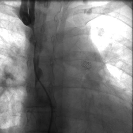
If a patient presently acutely with airway obstruction, the airway must be secured initially via intubation or surgical methods. After the patient has been stabilized, radiotherapy and corticosteroids should be considered. Urgent treatment with radiotherapy and corticosteroids should only be used for life-threatening situations and should be deferred otherwise due to interference with subsequent histopathological diagnosis. A useful procedure for patients with severe SVCS in respiratory distress is endovascular therapy with stenting; meta-analysis show it has high technical and clinical success rates. It is done via the femoral vein and performed under conscious sedation; fluoroscopic guidance and iodinated contrast are used alongside heparin. The image below shows SVC stenosis with stent placement in the left pulmonary artery.
Prognosis
Prognosis depends on underlying aetiology with poorer prognosis for malignant conditions. In malignancy, SVCS does not necessarily signify adverse outcomes. However, in patients with non-small cell lung cancer resistant to chemotherapy and radiotherapy, development of SVC syndrome is associated with poor prognosis and median survival of <6 months. In benign aetiologies, stenting or surgery has a patency rate of 90% although recurrent stenting may be required in some cases. Patient may need to be on antiplatelet therapy or warfarin for 1 to 3 months although there are no clear guidelines regarding the duration of treatment.
Differential diagnosis
Cardiac tamponade: Absence of facial and upper-extremity oedema with prominent x-descent in JVP
Constrictive pericarditis
Acute COPD exacerbation
Right sided heart failure
PE
Cardiac tumour