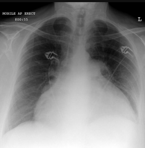
Chest X-Ray (CXR)
Systematic approach to look at the heart - ABCDEF (Radiopaedia)
When looking at a CXR, we can use ABCDEF to look at each area of the CXR:
A - Airways (intraluminal mass, narrowing, splayed carina)
Start at the top in the midline and review the airways:
Trace the trachea down to the carina and main bronchi
The trachea should be midline at the sternal notch, and deviates to the right around the aortic arch and divides into the right and left main bronchi with an angle less than 105' (mean 80')
Any narrowing or intraluminal lesions?
Trace down both main bronchi
Is the carina wide (more than 105 degrees?)
Is there bronchial narrowing or cut-off?
Is there any inhaled foreign body?
B - Breathing (lungs, pulmonary vessels, pleural spaces)
Both lungs should be well expanded and similar in volume
Can you count 10 posterior ribs bilaterally?
Is one lung larger than the other
Compare the apical, upper, middle and lower zones in turn
Are they symmetrical?
Are there areas of increased density
Trace the lung vessels
Can you see the vasculature equally throughout both lungs?
Can you see the retrocardiac and retro diaphragmatic lung vessels?
Are there extra lines in the periphery that aren't vessels?
Trace the lateral margins of the lung to the costophrenic angles
Are the costophrenic angles crisp?
Trace the hemidiaphragms to the vertebrae
Can you see the whole of the hemidiaphragm?
Trace the cardiac borders
Can you clearly see the left and right heart borders?
Can you see the descending aorta
Check the heart shadow for retrocardiac lung opacity
Check the diaphragm for overlying lung lesions in the posterior costophrenic recesses
C - Circulation (cardiomediastinal contour, great vessels)
Check the cardiac position
Is 1/3 to the right and 2/3 to the left?
Assess cardiac size
Is the cardiothoracic ratio < 50%?
Check the position and size of the aortic arch and pulmonary trunk
Check the width of the upper mediastinum
Look at the hilar vessels
Can you see them clearly on both sides?
Are they at a similar height?
Can you see a preserved hilar point bilaterally and a little higher on the left?
D - Diaphragm and below
Is the right hemidiaphragm the same height or up to 2cm higher then the left hemidiaphragm?
Is there a hiatus hernia?
Can you identify the gastric bubble, splenic flexure of the colon and spleen
Is there any free intraperitoneal gas?
Is the stomach or bowel dilated?
Are there any gallbladder or renal calculi?
E - External (chest wall, shoulder girdles)
Equal companion shadows to both clavicles
Symmetrical or left slightly lower breast shadows?
Soft tissue emphysema?
Trace along each posterior (horizontal) rib on one side the chest
Is there a fracture or area of destruction
Repeat with the other side of the chest
Trace lateral and anterior ribs on the first side
Repeat on the other side
Check the clavicles and shoulder girdles for bone destruction or dislocation
Can you trace around the cortex of the bones?
Check the vertebral bodies; at each level you should see two eyes (pedicles), a mouth (interlaminar space) and a nose (spinous process):
Are the bodies rectangular and of a similar height?
Can you see 2 pedicles per vertebral body?
Are there disc spaces?
F - Foreign material (devices, foreign bodies, gossypiboma)
Review the upper abdomen, soft tissues and chest
Are there any surgical clips?
Are there any devices?
Are lines and tubes in a satisfactory position
Are there any unexpected foreign bodies such as retained swabs?
Detailed approach and anatomy of the CXR
Mediastinum
Heart
The heart must be assessed for its shape, size and location. Abnormal cardiac shift may reflect ipsilateral loss volume, lobar atelectasis or contralateral increase volume (large pneumothorax).
Enlargement of the cardiac silhouette may be due to cardiomegaly or pericardial effusion. When large, pericardial effusion may result in a 'water bottle heart' sign as shown below.
The pericardial fat pad sign is formed by visualization of a pericardial effusion as a curvilinear band of soft tissue >2mm thick outlined by mediastinal fat anteriorly and subepicardial fat posteriorly.
Cardiac calcifications may correspond to a coronary artery, valvular or annular c calcifications, or curvilinear calcification in a left ventricular aneurysm from prior MI.