Visual field defects
A careful analysis of the visual field defect can often indicate the site of neurological damage - relatively large visual field deficits are called anopsias while smaller ones are referred to as scotomas.
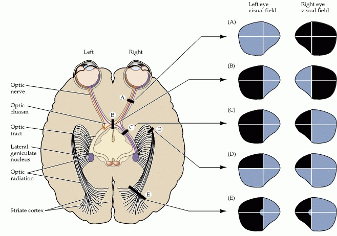
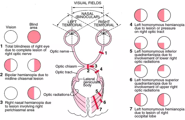
Damage to the deeper brain structures responsible for vision, such as the optic tract, lateral geniculate nucleus or optic radiation and visual cortex, will result in issues that are limited to the contralateral visual hemifield - such as seen in number 4 and 7 and letter C.
Damage to the optic chasm results in visual field deficits that involve non-corresponding parts of the visual field in each eye. This is often the result of pituitary tumours. It leaves the uncrossed portion of each fibre intact, which causes the temporal region (which would be the nasal region of sight) to not be affected. It is known as a bitemporal hemianopsia as the temporal areas that the optic nerve supplies is affected, although the side of sight affected is nasally. This is seen in number 2 and letter B.
Damage to the central visual structures is rarely complete - as a result, the deficits associated with damage to the chaism, optic tract/radiation, or visual cortex are typically more limited then shown in the diagrams above. This is especially true for damage along the optic radiation, which fans out under the temporal and parietal lobes in the course from the lateral geniculate nucleus to the striate cortex. Some of the optic radiation axons run out into the temporal lobe on their path to the striate cortex, an anomaly called Meyer's loop. Damage to parts of the temporal lobe with involvement of Meyer's loop can thus result in a superior homonymous quadrantanopsia. It will be a contralateral (if the right optic radiation is damaged, this will reflect in a left superior homonymous quadrantanopsia. If the lower radiation is damaged, this will be a lower superior homonymous quadrantanopsia. If the upper is damaged, it will be an upper.
Injury to central visual structures can also lead to a phenomenon called macular sparing - loss of vision throughout wide areas with the excpetion of foveal vision. It is commonly found in damage to the visual cortex. The explanaition for macular sparing is not clear - it may be due to overlap in the pattern of crossed and uncrossed ganglion cells supplying central vision.