Parathyroid and the axis/role of Parathyroid Hormone
Parathyroid gland
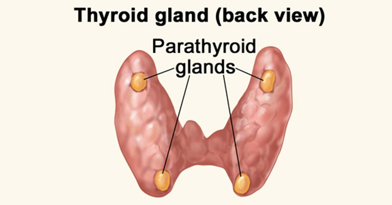
The parathyroid is comprised of 4 small glands which are embedded in the posterior aspect of the thyroid glands. The primary function is the production and secretion of parathyroid hormone (PTH), polypeptide hormone responsible for maintaining serum calcium homeostasis.
The levels of PTH and serum calcium are inversely proportional - the higher the levels of PTH, the lower the levels of serum calcium. At low serum calcium, PTH, in conjunction with vitamin D, works at many sites in the body to mobilise calcium stores and increase absorption and reabsorption. Both calcium and vitamin D provide negative feedback to the parathyroid glands; as calcium and vitamin D levels increase, they bind receptors at the parathyroid gland and inhibit the production and release of PTH.
Cellular level
Chief cells
The chief cells are the functional cells of the parathyroid gland - responsible for both synthesising and secreting parathyroid hormone. Regulation of PTH production and release is dependent on serum calcium levels.
The G-protein coupled transmembrane receptor, calcium-sensing receptor (CaSR), on the surface of chief cells respond to low serum calcium and activates translation and secretion of PTH
Oxyphil cells
Also known as oxyntic cells - they have no recognised endocrine function. The proportion of oxyphil cells in the parathyroid glands increases with age.
Development
The 4 parathyroid glands develop from the endoderm of the 3 and 4 pharyngeal pouches around 6 weeks of gestation. Studies have suggested that there may be some ectodermal and neural crest contribution of the glands formation as well.
The inferior parathyroid glands develop from the 3 pharyngeal pouches, while the superior glands develop from the 4 pharyngeal pouches. This is due to the additional formation of the thymus by these pharyngeal pouches that migrate caudally later in gestation.
Parathyroid glands are functional during gestation, acting to control calcium balance in the fetus.
Function
The parathyroid glands function is to maintain serum calcium homeostasis through the synthesis and release of PTH. At the bone, PTH inhibits osteoblast activity and stimulates osteoclast activity leading to bone breakdown and calcium release.
PTH increases calcium reabsorption at the kidneys and block phosphate reabsorption from the tubules. PTH also acts on the kidneys to stimulate the formation of vitamin D. Vitamin D is an essential component of calcium and phosphate homeostasis, with its effects on the kidneys and GI system.
Skeletal system
PTH works at the cellular levels by indirectly stimulating osteoclasts to break down bone. PTH binds to cell receptors on osteoblasts stimulating the release of Receptor Activator of Nuclear Factor Kappa-B ligand (RANKL). RANKL binds to its receptor on osteoclast precursors, stimulating them to fuse into mature osteoclasts, thereby increased calcium resorption from the bone.
Renal system
PTH has 2 main roles in the kidney. It works in two places:
DCT
Collecting ducts to increase reabsorption of calcium by up-regulating TRPV5 (a calcium transporter on the tubular epithelium)
PTH also binds to sites in the proximal tubule that block phosphate reabsorption. Thus, the net effect of PTH is to decrease calcium excretion and increase phosphate excretion in the urine. PTH works at the proximal tubule of the kidneys to up-regulate the translation of alpha-1-hydroxylase, the enzyme responsible for generating the biologically active form of vitamin D (1,25-dihydroxy-vitamin D or calcitriol).
Vitamin D
Vitamin D binds to receptors in the bone that function similarly to PTH, increasing serum calcium. In the kidney, vitamin D increases both calcium and phosphate reabsorption.
Gastrointestinal
Calcium absorption occurs in the small intestines, with 70% to 80% absorbed in the ileum. Although PTH has no direct effects on the small intestine, the downstream effects of PTH on vitamin D synthesis occur at this level. Vitamin D increases calcium and phosphate absorption from the gut. There are vitamin D receptors across the entirety of the gut epithelium, and studies have shown both paracellular and transcellular absorption of calcium.
VItamin D also increases transcription and translation of calcium transport proteins in the epithelium including TRPV6, calbindin and ATP-dependent calcium pumps
Testing
Total Serum Calcium
Total serum calcium is the amount of calcium in the blood including free or ionised, complexed calcium (calcium bound to bicarbonate or citrate), and protein-bound calcium. Normal levels are between 8.5 to 10.2mg/dL [0.85 and 1.2 umg/dL]. The value is affected by amount of protein, usually albumin, available for binding and thus must be corrected in hypoalbuminemia. For every 1 gm decrease in serum albumin, the total serum calcium decreased by approximately 0.8mg. The offical equation is:
Corrected calcium = [Ca + 0.02 x (40 - albumin] where Ca is in mg/dL
Ionised serum calcium
Ionized calcium (free calcium) is the biologically active form of calcium needed for many functions such as cell signaling, neurotransmission, and muscle contraction. Normal levels range from 4.64 to 5.28 mg/dL. Blood pH can affect ionised calcium levels by changing the binding affinity of albumin for positively charged anions. At a low pH, an abundance of hydrogen ions effectively competes with free calcium for binding sites on negatively charged albumin, thus increasing free calcium in the serum.
Parathyroid hormone
PTH levels can be measured through immunoassays. Normal levels range from 10 to 65 pg/mL.
Parathyroid-related peptide
PTHrP is a peptide produced by nearly all tissues in the body at very low levels. The physiologic function is unknown, but it appears to serve mostly autocrine and paracrine functions, accounting for its low plasma level. Normal levels are < 2.0 pmol/L. PTHrP is overproduced in a wide variety of carcinomas but is most notably associated with squamous cell lung cancer, squamous cell cancers of the head and neck, and breast cancer. Levels of PTHrP greater than the reference range warrant a thorough searched for related cancers.
Sestamibi parathyroid scintigraphy
A sestamibi parathyroid scan is ordered in patients who are believed to have parathyroid adenomas or hyperplasia - the radioisotope technetium-99m is injected IV and taken up by active oxyphil cells in the parathyroid tissue.
A gamma camera is used to take images of the neck and will show the locations and sizes of the parathyroid glands. Repeat images are taken after a washout periods of 2 to 4 hours, and glands still showing activity are considered hyperactive.
Vitamin D
Most assay measuring vitamin D test the biologically inactive form 25-OH as it is the major form of vitamin D in circulation. As the active metabolite, 1-25-OH, has a circulating half life of 4 to 6 hours, making it difficult to interpret. Normal vitamin D levels range from 30 to 100. Between 21 to 29 suggests insufficiency and levels below 20 suggest deficiency.